Ct Anatomy Pelvis Muscles - Abdomen And Pelvis Ct : Ct of the abdomen axial anatomy.. If you want to learn how to read ct scans of the abdomen and pelvis proficiently, this video is an excellent starting point. Free and interactive atlas of the human anatomy. Microscopic anatomy of skeletal muscle. The pelvis is a developmentally complex skeletal structure requiring the fusion of separate elements and articulation with both the axial skeleton and lower limb. Involved early gray = muscle:
How to view anatomical labels. Related online courses on physioplus. These muscles, including the gluteus maximus and the hamstrings, extend the thigh at the hip in support of the body's weight and propulsion. Key facts about the muscles of the pelvic floor. Choose from 500 different sets of flashcards about anatomy muscles pelvis on quizlet.
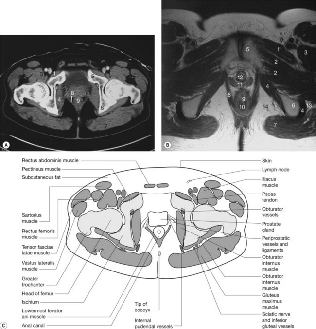
Various Approaches For Ct Guided Percutaneous Biopsy Of Deep Pelvic Lesions Anatomic And Technical Considerations Radiographics from pubs.rsna.org Migdalia ordonez ohsu summer 2012. In the back the posterior superior iliac spines are surrounded by muscles and flank fat. The full bladder displaces small bowel loops superiorly. This is a normal view of the pelvis. Innervation of the female levator ani muscles. Figure 6.4 • ct scan of pelvis: Spin it around and draw the bucket! It is a powerful hip extensor that acts to bring the thigh in a straight line with the pelvis.
If you want to learn how to read ct scans of the abdomen and pelvis proficiently, this video is an excellent starting point.
Normal ct abdomen/pelvis (without labels). The gluteus maximus is a superficial muscle of the hip that forms most of the flesh of the buttock; Other pelvic muscles, such as the psoas major and iliacus, serve as flexors. Included within the chart are gorgeous illustrations of the pelvic diaphragm, sphincter muscles, gluteus maximus. Anatomy of the muscular system. Some of the most important include the major digestive organs, the intestines. Spin it around and draw the bucket! Functional anatomy of the male. This is the iliopubic line which outlines the anatomic anterior column this is the. The pelvis is a developmentally complex skeletal structure requiring the fusion of separate elements and articulation with both the axial skeleton and lower limb. If you want to learn how to read ct scans of the abdomen and pelvis proficiently, this video is an excellent starting point. Anatomy of the abdominal cavity and the male pelvis: Attached to the pelvis are muscles of the buttocks, the lower back, and the thighs.
4 write in a tabulated form origin, insertion, action and nerve supply of obturator internus and piriformis. 3 enumerate the muscles of true pelvis. If you want to learn how to read ct scans of the abdomen and pelvis proficiently, this video is an excellent starting point. Pelvic floor muscles that are located wholly within the pelvis. This mri male pelvis axial cross sectional anatomy tool is absolutely free to use.
The Pelvis Radiology Key from radiologykey.com Anatomy of the abdominal cavity and the male pelvis: This is a normal view of the pelvis. Axial section through male bladder. Spin it around and draw the bucket! The muscles of the pelvis, hip and buttock anatomical chart shows how each muscle in this area of the body works with the others, and the various minor systems within the major ones. Migdalia ordonez ohsu summer 2012. Ct abdomen ct pelvis / 5. How to view anatomical labels.
This is a normal view of the pelvis.
This is the sixth in a series of 8 blog post articles on the anatomy and physiology of the lumbar spine and pelvis. Free and interactive atlas of the human anatomy. Key facts about the muscles of the pelvic floor. Ct of the abdomen axial anatomy. Innervation of the female levator ani muscles. When looking for acetabular fractures there a few lines to look at. If you want to learn how to read ct scans of the abdomen and pelvis proficiently, this video is an excellent starting point. The muscles of the pelvis, hip and buttock anatomical chart shows how each muscle in this area of the body works with the others, and the various minor systems within the major ones. Migdalia ordonez ohsu summer 2012. Axial section through male bladder. Learn about anatomy muscles pelvis with free interactive flashcards. Muscles of the pelvis that cross the lumbosacral joint to attach onto the trunk were described in the previous blog post note: The gluteus maximus is a superficial muscle of the hip that forms most of the flesh of the buttock;
Functional anatomy of the male. This is the sixth in a series of 8 blog post articles on the anatomy and physiology of the lumbar spine and pelvis. Use the mouse scroll wheel to move the images up and down alternatively use the tiny arrows (>>) on both side of the image to move the images. This is a table of skeletal muscles of the human anatomy. This mri male pelvis axial cross sectional anatomy tool is absolutely free to use.
Gi And Abdomen Radiologic Anatomy from www.meddean.luc.edu This page provides a photo gallery that presents the anatomy of the abdomen by means of ct (axial, coronal, and sagittal reconstructions). Ct abdomen ct pelvis / 5. This tool provides access to a ct atlas in the axial plane, allowing the user. Anatomy of the muscular system. The small intestine is the longest part of the digestive tract. It is a powerful hip extensor that acts to bring the thigh in a straight line with the pelvis. In the back the posterior superior iliac spines are surrounded by muscles and flank fat. Some of the most important include the major digestive organs, the intestines.
The muscles of the pelvis form its floor.
This is a normal view of the pelvis. Figure 6.4 • ct scan of pelvis: Axial section through male bladder. Ct of the abdomen axial anatomy. There are many muscles that form the pelvic floor, including puborectalis, pubococcygeus, iliococcygeus and coccygeus. Pelvic examinations are common in clinical cases of obstetrics and gynecology the bony pelvis can be divided and viewed into 2 parts: Architectural differences in the bony pelvis of women with and without pelvic floor disorders. Related online courses on physioplus. Some of the most important include the major digestive organs, the intestines. The full bladder displaces small bowel loops superiorly. The muscles of the pelvis form its floor. Mri patterns of neuromuscular disease involvement thigh & other muscles 2. The small intestine is the longest part of the digestive tract.
Furthermore, the pelvis protects the pelvic and abdominopelvic viscera anatomy muscles pelvis. Architectural differences in the bony pelvis of women with and without pelvic floor disorders.